Neck problem, Pain and discomfort

Common neck pain:

  • Osteoarthritis (worn joints) in neck tends to occur with age
  • Neck muscle strained because poor posture and overuse. For example prolonged driving, reading in bed
  • Whiplash – where there is a sudden jolt to the head and neck that is beyond the normal range of the motion
  • Herniated Disc or a pinched nerve: A herniated (slipped) disk occurs when all or part of a disk in the spine is forced through a weakened part of the disk. This places pressure on nearby nerves.
  • Torticollis: Torticollis means ‘twisted neck’.
  • Other diseases like Cancer, Rheumatoid Arthritis, Meningitis

What is Neck Pain?

Pain located in the neck usually at the back or sides.

The neck (cervical spine) is composed of vertebrae that begin in the upper torso and end at the base of the skull. The bony vertebrae along with the ligaments (which are comparable to thick rubber bands) provide stability to the spine. The muscles allow for support and motion. The neck has a significant amount of motion and supports the weight of the head. However, because it is less protected than the rest of the spine, the neck can be vulnerable to injury and disorders that produce pain and restrict motion. For many people, neck pain is a temporary condition that disappears with time. Others need medical diagnosis and treatment to relieve their symptoms.

What are the signs and symptoms of Neck Pain?

  • General pain located in the neck area, as well as stiffness in the neck muscles.
  • The pain may radiate down to the shoulder or between the shoulder blades.
  • It may also radiate out into the arm, the hand or up into the head, causing a one-sided or double-sided headache.
  • The muscles in the neck are tense, sore and feel hard to the touch.
  • Acute pain can give rise to abnormal neck posture in which the head is forced to turn to one side. This condition is known as torticollis.
  • The pain at the base of the skull may be accompanied by a feeling of weakness in the shoulders and arms.
  • There may be a prickly or tingling sensation in the arms and fingers.

How is Neck Pain evaluated?

An examination of the nervous system is performed to determine whether nerve involvement is present. Further testing includes x-ray evaluation, CAT scan, bone scan and MRI scan.

What are the causes of Neck Pain?

Neck pain may result from abnormalities in the soft tissues—the muscles, ligaments, and nerves—as well as in bones and joints of the spine. The most common causes of neck pain are soft-tissue abnormalities due to injury or prolonged wear and tear. In rare instances, infection or tumors may cause neck pain. In some people, neck problems may be the source of pain in the upper back, shoulders, or arms.

Many things can trigger neck pain. These include:

  • trauma or injury
  • worry and stress
  • falling asleep in an awkward position
  • prolonged use of a computer keyboard.

What are the treatments for Neck Pain?

The treatment of neck pain depends on its precise cause. Treatment options include:

  • Rest
  • Heat / Ice applications
  • Traction
  • Soft Collar
  • Physical therapy (ultrasound, massage, manipulation)
  • Local injections of cortisone or anesthetics
  • Topical anesthetic creams
  • Topical pain patches
  • Muscle Relaxants
  • Analgesics
  • Surgical procedures may require for severe case

Neck pain with pain radiating to arm

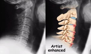
X-ray showing bone spur

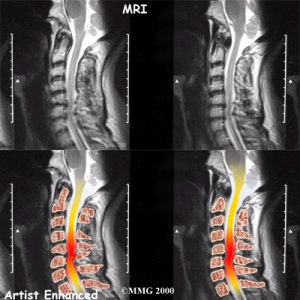
MRI showing prolapsed cervical disc

NOTE: If severe neck pain occurs following an injury (motor vehicle accident, diving accident, or fall), a trained professional, like our senior orthopeadic surgeon Dr Kevin Yip Man Hing should immobilize the patient to avoid the risk of further injury and possible paralysis.

For more information or an appointment with our senior orthopeadic surgeon Dr Kevin Yip Man Hing, please dial our 24HR hotline at (+65) 6471 2691

A must to know, Back pain

Do you ever have a sleepless night due to back pain? or after long hour or work, in office, or sports activities and you encounter back pain?

Below are some of the symptoms that caused back pain.

  • Slip Disc / Herniated Disc
  • Scoliosis
  • Sciatica
  • Tail Bone (Coccydynia)
  • Spondylolisthesis
  • Mid Back Pain (Thoracic Pain)
  • Back Strain
  • Lower Back Pain
It’s time to care for your back. Don’t let back pain affect the quality of your life! Simply call us for an appointment Now!

Do you or anyone around you ever complain of backache? Backache or back pain is a common disorder which affects the quality of life of many. Back pain can be short in duration or sometimes persistent. In most cases, the pain subsides on its own without treatment within a few weeks. However, not every back pain can be self-treated. If you have back pain that does not improve after weeks, backache associated with numbness or tingling, severe pain that does not improve after rest, and pain after a fall or an injury, you should see doctor immediately by calling us for an appointment.

Symptoms of Back Pain

  • Muscle ache
  • Pain that radiates down your leg
  • Numbness or weakness in one or both legs or around your buttock
  • Pain that is getting worse and is spreading up your spine like shooting or stabbing pain
  • Inability to stand straight
  • Limited range of movement of your back

Causes of Back Pain

  • Aging. As we grow older, our spine ages with us too, especially after 30 years old.
  • Physically inactive lifestyle. Sitting for long hours without proper posture and prolonged standing may lead to back problems. So, do you spend more time in from of the desk working or playing games or watching TV for long hours? Do you often lean back to relax or sliding down on your favorite sofa with bad posture? If your answer is yes to one or more of those bad-straining habits, it won’t do any good for your back in the long run.
  • Sleeping position. Unhealthy sleeping position can aggravate your back problem.
  • Obesity. People with obesity have added force on their lower back and aggravate the pain.
  • Fracture – a crack or break in one of the bones in your back
  • Other diseases. Those with existing back conditions such as slipped discs, arthritis in the spine, osteoporosis and others are at higher risk of having persistent back pain.

How’s Back Pain diagnosed?

  • X-ray
  • MRI

Back Pain Solutions

  • Medications like anti-inflammatory medication, pain relieves, and nerve regenerating medicatio
  • Hot / Cold packs can apply to the affected area in order to help to relieve the pain temporarily by soothing sore and stiff back.
  • Back Supports for better comfort leve
  • Physiotherapy. Treatment can involve exercises, posture advice, massage, and techniques known as spinal mobilization and spinal manipulation. Treatment course normally last about 6 to 12 weeks.
  • Epidural Injection. This type of injection can be done at Clinic as outpatient treatment
  • Surgery may consider for severe cases when the back pain does not improve after several attempts to conservative treatments. On average, about 1 in 10 people may require the surgery and surgery is considered to be last resort of treating the chronic back pain. Fenestration and Discectomy may require for prolapsed intervetebral disc.

How to prevent back pain?

  • Find your correct standing posture by squeezing you buttocks, pulling your “pits to your hips”, and tucking in your chin
  • Take frequent standing breaks top bend backward 5-6 repetitions, every hour if you have low back pain
  • Find your correct sitting posture using the slouch-overcorrect technique. Do 10 repetitions, 3 times a day, and at any time you experience pain.
  • Avoid twisting your body when moving an object
  • Lift with your legs keeping your back straight, standing as close as possible to the object to be lifted
  • Take regular exercise like swimming


  • Good sleeping postures
    If you are a side sleeper, draw your legs up slightly towards your chest and put a pillow between your legs.
    If you are a back sleeper, placing pillow under your knees will help to maintain the normal curve of your back.


  • Occupational – related. Back pain is “collar-blind”, affecting both blue and white-collar workers. However, blue-collar workers are more prone to having more back problems than white-collar workers. Bending, lifting, pushing, twisting or straining too harsh can result in back pain.

Shoulder pain, how does it occur?

Do you or anyone around you ever complain of Shoulder Pain?

Shoulder pain is a common disorder which affects the quality of life of many. Back pain can be short in duration or sometimes persistent. In most cases, the pain subsides on its own without treatment within a few weeks. However, not every shoulder pain can be self-treated. If you have shoulder pain that does not improve after weeks, shoulder ache associated with numbness or tingling, severe pain that does not improve after rest, and pain after a fall or an injury, you should see doctor immediately or call us to make an appointment at +65 6471 2691

Anatomy of Shoulder

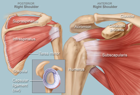
The shoulder is one of the largest and most complex joints in the body. The shoulder joint is formed where the humerus (upper arm bone) fits into the scapula (shoulder blade), like a ball and socket. Other important bones in the shoulder include:

  • The acromion is a bony projection off the scapula.
  • The clavicle (collarbone) meets the acromion in the acromioclavicular joint.
  • The coracoid process is a hook-like bony projection from the scapula.

The shoulder has several other important structures:

  • The rotator cuff is a collection of muscles and tendons that surround the shoulder, giving it support and allowing a wide range of motion.
  • The bursa is a small sac of fluid that cushions and protects the tendons of the rotator cuff.
  • A cuff of cartilage called the labrum forms a cup for the ball-like head of the humerus to fit into.

The humerus fits relatively loosely into the shoulder joint. This gives the shoulder a wide range of motion, but also makes it vulnerable to injury.

Introduction of Shoulder Pain

Shoulder pain includes any pain that arises in or around your shoulder. Shoulder pain may originate in the joint itself, or from any of the many surrounding muscles, ligaments or tendons. Shoulder pain usually worsens with activities or movement of your arm or shoulder.

The shoulder is the most mobile joint in the human body. A group of four tendons in the shoulder, called the rotator cuff, give the shoulder a wide range of motion.

Swelling, damage, or bone changes around the rotator cuff can cause shoulder pain. You may have pain when lifting the arm above your head or moving it forward or behind the back.

Symptoms of Shoulder Pain

  • Swelling
  • Redness
  • Tenderness and warmth around the joint
  • Limited Range of movement

What causes Shoulder Pain?

  • Rotator Cuff Tendonitis – the most common cause of shoulder pain – when rotator cuff tendons become trapped under the bony area in the shoulder. The tendons become inflamed or damaged.
  • Fracture shoulder bone, e.g. Collar Bone
  • Frozen shoulder, which occurs when the muscles, tendons, and ligaments inside the shoulder become stiff, making movement difficult and painful
  • Overuse or injury of nearby tendons, such as the bicep muscles of your arms
  • Dislocation of your shoulder
  • Bursitis is Inflammation of a fluid-filled sac (bursa) that normally protects the joint and helps it move smoothly
  • Arthritis in the shoulder joint

Sometimes, shoulder pain may be due to a problem in another area of the body, such as the neck or lungs. This is called “referred pain.” People with this type of pain usually do not have pain when moving the shoulder.

How is Shoulder Pain diagnosed?

  • X-rays: Plain X-rays can reveal narrowing of the space between two spinal bones, arthritis-like diseases, tumors, slipped discs, narrowing of the spinal canal, fractures and instability of the spinal column.
  • MRI: Magnetic Resonance Imaging is a noninvasive procedure that can reveal the detail of neural (nerve-related) elements.

Treatment for Shoulder Pain

All treatments will be varied on individual depending on the source of pain.

Treatment for shoulder pain may include:

  • Nonsteroidal anti-inflammatory drugs (NSAIDs)
  • Injection of a powerful anti-inflammatory medicine called a corticosteroid
  • Physical therapy
  • Surgery if all other treatments do not work

Prevention for Shoulder Pain

  • If you have had shoulder pain before, use ice and take anti-inflammatory medicine after exercising.
  • Learn proper exercises to stretch and strengthen your rotator cuff tendons and shoulder muscles. A doctor or physical therapist can help.
  • If you are recovering from tendinitis, continue to perform range-of-motion exercises to avoid “frozen shoulder.”

For more information, please call (65) 6471 2691

Knee Pain, What is it?

Do you or anyone around you ever complain of knee pain?

Knee pain is a common disorder which affects the quality of life of many and affects to all ages. Knee pain can be short in duration or sometimes persistent. In most cases, the pain subsides on its own without treatment within a few weeks. If you have knee pain that does not improve after weeks, you should see doctor immediately or you can call us at +65 6471 2691. Delays in diagnosis and treatment can lead to poor healing, reduced range of motion and long-term disability.

Anatomy of Knee

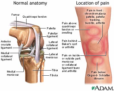
The knee joint is the largest joint in the body, consisting of 4 bones and an extensive network of ligaments and muscles. Injuries to the knee joint are amongst the most common in sporting activities and understanding the anatomy of the joint is fundamental in understanding any subsequent pathology.

The knee is made up of:

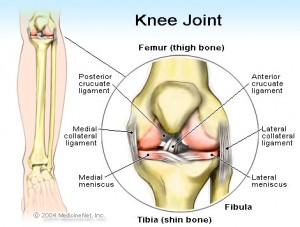
  • Four main Bones- the femur (thigh bone), the tibia (shin bone), fibula (outer shin bone) and patella (kneecap).
  • Joint capsule: a thick ligamentous structure that surrounds the entire knee.
  • Four Ligaments- Anterior Cruciate Ligament (ACL), Medial Collateral Ligament (MCL), Lateral Collateral Ligament (LCL), Posterior Cruciate Ligament (PCL)
      • Medial collateral ligament (MCL) runs along the inside of the knee and prevents the knee from bending out
      • Lateral collateral ligament (LCL) runs along the outside of the knee and prevents the knee from bending in
      • Anterior cruciate ligament (ACL) is in the middle of the knee. It prevents the shin bone from sliding out in front of the thigh bone
      • Posterior cruciate ligament (PCL) works with the ACL. It prevents the shin bone from sliding backwards under the femur
  • Meniscus: Each knee joint has two crescent-shaped cartilage menisci. These lie on the medial (inner) and lateral (outer) edges of the upper surface of the tibia bone
  • The two main Muscle groups of the knee knee joint are the quadriceps and the hamstrings.

Common Knee Pains

  • Knee pain due to meniscus injury
    The meniscus is a small “c” shaped cartilage that acts as a cushion in the knee join. The meniscus sit between the femur and the tibia bone, one on the outside and one on the inside of the knee. A meniscus tear occurs when the cartilage tear and are injured usually during movement that forcefully rotate the knee while weight bearing.
  • Knee pain due to osteoarthritis
    Knee pain due to osteoarthritis is a degenerative disease that results in a gradual wearing away of joint cartilage. As we grow older, we tends to get this problem.
  • Knee pain due to ligament torn
    Knee ligaments help to keep your knee stable. Four ligaments that made up of a knee are – Anterior Cruciate Ligament (ACL), Medial Collateral Ligament (MCL), Lateral Collateral Ligament (LCL), Posterior Cruciate Ligament (PCL)
  • Baker’s cyst
    an accumulation of joint fluid (synovial fluid) that forms behind the knee.
  • Patellar Quadriceps Tendonitis
    is also known as “Jumper Knee”. Patellar tendonitis is a common overuse injury. It occurs when repeated stress is placed on the patellar tendon. The stress results in tiny in the tendon, which the body attempts to repair. However when the tears in the tendon increase faster than what the body can recover, it cause the inflammation in the tendon to worsen.
  • Plica Syndrome
    Often called “synovial plica syndrome,” this is a condition that is the result of a remnant of fetal tissue in the knee. The synovial plica are membranes that separate the knee into compartments during fetal development. These plica normally diminish in size during the second trimester of fetal development. In adults, they exist as sleeves of tissue called “synovial folds,” or plica. In some individuals, the synovial plica is more prominent and prone to irritation.
  • Osteochondritis dissecans
    is a joint condition in which a piece of cartilage, along with a thin layer of the bone beneath it, comes loose from the end of a bone.
  • Chondromalacia patellae (patellofemoral pain syndrome)
    general term that refers to pain arising between your patella and the underlying thighbone (femur). It’s common in young adults, especially those who have a slight misalignment of the kneecap; in athletes; and in older adults, who usually develop the condition as a result of arthritis of the kneecap.
  • Osgood-Schlatter disease
    an overuse injury that occurs in the knee area of growing adolescents. It is caused by inflammation of the tendon below the kneecap (patellar tendon) where it attaches to the shinbone (tibia).
  • Gout
    a disease that involves the build-up of uric acid in the body that can affect joints in your body. About 95 percent of gout patients are men. Most men are over 50 when gout first appears. Women generally don’t develop gout until after menopause. But some people develop gout at a young age.
  • Knee Strain - cause by violent stretching of one or more ligaments in the knee. Sprains involving two or more ligaments cause considerably more disability than single-ligament sprains.

Symptoms of knee pain

The location and severity of knee pain may vary, depending on the cause of the problem. Signs and symptoms that sometimes accompany knee pain include:

  • Swelling and stiffness
  • Redness and warmth to the touch
  • Weakness or instability
  • Popping or crunching noises
  • “Locking,” or unable to fully extend

Common Treatments for Knee Pain

Treatments will vary, depending upon what exactly is causing your knee pain.

Medications

Your doctor may prescribe medications to help relieve pain and to treat underlying conditions, such as rheumatoid arthritis or gout.

Therapy

  • Physical therapy. Strengthening the muscles around your knee will make it more stable. Training is likely to focus on the muscles on the front of your thigh (quadriceps) and the muscles in the back of your thigh (hamstrings). Exercises to improve your balance are also important.
  • Orthotics and bracing. Arch supports, sometimes with wedges on the inner or outer aspect of the heel, can help to shift pressure away from the side of the knee most affected by osteoarthritis. Different types of braces may help protect and support the knee joint.

Injections

  • Corticosteroids. Injections of a corticosteroid drug into your knee joint may help reduce the symptoms of an arthritis flare and provide pain relief that lasts a few months. The injections aren’t effective in all cases. There is a small risk of infection.
  • Hyaluronic acid. This thick fluid is normally found in healthy joints, and injecting it into damaged ones may ease pain and provide lubrication. Experts aren’t quite sure how hyaluronic acid works, but it may reduce inflammation. Relief from a series of shots may last as long as six months to a year.

Surgery

If you have an injury that may require surgery, it’s usually not necessary to have the operation immediately. Before making any decision, consider the pros and cons of both nonsurgical rehabilitation and surgical reconstruction in relation to what’s most important to you. If you choose to have surgery, your options may include:

  • Arthroscopic surgery. Depending on the nature of your injury, your doctor may be able to examine and repair your joint damage using a fiber-optic camera and long, narrow tools inserted through just a few small incisions around your knee. Arthroscopy may be used to remove loose bodies from your knee joint, repair torn or damaged cartilage and reconstruct torn ligaments.
  • Partial knee replacement surgery. In this procedure (unicompartmental arthroplasty), your surgeon replaces only the most damaged portion of your knee with parts made of metal and plastic. The surgery can usually be performed with a small incision, and your hospital stay is typically just one night. You’re also likely to heal more quickly than you are with surgery to replace your entire knee.
  • Total knee replacement. In this procedure, your surgeon cuts away damaged bone and cartilage from your thighbone, shinbone and kneecap, and replaces it with an artificial joint made of metal alloys, high-grade plastics and polymers.
For more information, please call (65) 6471 2691 – 24HR HOTLINE

Treatment For Scoliosis

SCOLIOSIS is a spinal abnormality in normal condition straight transformed into the form of the letter “S”.
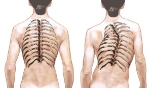
Generally the patient is never aware of this disorder and he only realized after notified by the family or close relatives who accidentally see any difference. Some are only realized after his massage notified by builders who felt an unusual lump on his client’s body.

The presence of scoliosis will certainly have an impact on health is concerned which can start with the lightest such as pain, muscle pain, nerve pain, respiratory disruption, psychological disorders and even difficulty working because of limited motion in the cause.

Causes of Scoliosis

When viewed from the top 85% of the scoliosis is unknown or known as idiopathic, while the rest may be due to causes such as congenital disorders / birth defects, nervous system disorders, connective tissue, or other causes.

Asymmetric limb growth also widely referred to as the cause of scoliosis is why scoliosis is also known as structural or nonstructural abnormalities. The presence of factor descendants also quite large which is found in some cases of scoliosis in the family members

Diagnosis of Scoliosis

To detect or diagnose the presence of scoliosis do a physical examination by a physician experienced in handling cases of scoliosis and also in conjunction with radiological examinations done / X-ray.

With radiological examination the form of scoliosis that occurs can be easily identified. In addition it is also important to know besasrnya Cobb angle formed by the scoliosis condition. The angles formed by the arms of the spine will determine what kind of medical procedures undergone by the patient yangharus.

From the research that is then said to be formed when the Cobb angle less than 10 degrees then the least that should be done regularly so that regular observations can be minimized due to the possible. How to consult a physician who has handled before and radiology re-examination.

When the angle is between 10 to less than 40 degrees then the corresponding need of yangbersifat nonoperatif, but if the angle is more than 40 degrees, the patient requires assistance from a specialist orthopedic surgery fatherly / correction.

Another advantage of radiology examinations are pelvic examination can be done in order to set the level of development or fusion of the bone itself. When visible bone fusion is still not / do not merge the possibility of scoliosis patients worse becomes larger and also the opposite situation.

Sometimes patients feel reluctant to perform radiology examinations by various reasons and asked the doctor to do the examination at the next opportunity, or even at the end of treatment. For physician examination absolutely must be done so that he / doctor and the patient’s family can know the state of scoliosis in the early treatment of disease and the results of such an examination is very valuable because it will be used to compare the results of treatment with the initial state before handling the case.

Treatment For Scoliosis

Treatment or Management for Scoliosis doesn’t mean it will give the results of of a normal bone like 100% straight. This is considering that the bone is growing so that when it is found at an earlier age would provide a much better result than in adulthood.

Thus the aim of the treatment of the stretcher over to prevent damage to other organs caused by the occurrence of scoliosis. That means we still have to find the cause of scoliosis itself and wherever possible resolved that the disease does not progress to worse.

To handle as mentioned above may be operative and non-operative according to the lightness or severeness of scoliosis. For handling nonoperatif is currently offered so many advertisements that claim to cure scoliosis, but is actually just a medically recognized is the use of brace or support that serves as a buffer from the bone which is not straight. Scoliosis brace used on the course not the same as that used by fellow dentists to repair teeth arrangement because there scoliosis soft tissue such as skin is not as hard as the teeth and will also cause pain overemphasis pad. Brace therefore can not simply be bought, but must go through the patient’s condition. In addition, patients should also be disciplined in using it and most of his active life he had to use the brace. Constraints faced by patients at the time of use is a feeling of tightness, heaviness, heat and sometimes itchy sometimes causing patients not disciplined in use resulting in the ineffectiveness of the brace that ultimately can not deliver results as expected with.

Sports Therapy and Scoliosis

When patients experience difficulties in using the brace then do sports therapy is one way that can be used considering sports therapy is a treatment program that uses sports to measurably improve the health of a person or help cure certain diseases such as scoliosis. The use of sports therapy together is also a combination that can be done to improve the effectiveness of the undertaken treatment.

In cases of scoliosis will be an imbalance of working muscles in the spine to change shape and vice versa where the muscles that make the spine is tilted to one side. Thus we can give you a workout that will making working these muscles become more balanced and also resulted in a more straight spine, consequently the body becomes more relaxed and comfortable of course.

Exercises are performed will become more efficiently by using the computer equipment is referred to as EMG / Electrical MyoGraphy. In the presence of such a device would be seen as what types of exercises recruit the most muscle fibers during a workout which would impact on the acceleration of the results to be obtained by the patient concerned.

What also needs to be known is as therapy theray other sports do not make changes directly to the bone changes shape so that the repair is more temporary, which is why patients should continue to diligently perform the exercise has been known to be sustainable. It is equivalent to the brace that would otherwise be used again megembalikan original slant to forms bone. Therefore, when not practicing sports therapy regularly then auto-muscles are also no longer able to repair the abnormal bone structure.

This does not mean patients should absolutely continue to practice in sports therapy but just do exercises at home megingat he has been able to do it yourself, and after that to monitoring of the results of the exercise can be consulted on the treating physician. For sufferers \ scoliosis also be counting on weight because weight loss. Excessive especially for those who suffer from obesity will further aggravate the situation of his / her scoliosis.

Conclusion of Scoliosis

Scoliosis is a disorder that occurs in the spine that cause is still unclear, but if the cause is already made it known that the condition usually can also be resolved. In handling can besifat operative and nonoperatif. For those who nonoperatif overcome scoliosis with sports therapy is the best option because it is active in training the muscles are weak and can be combined with other nonoperatif treatment.

The result is although it will not be a normal people with scoliosis are forever, but at least it can reduce the negative impact caused by scoliosis and therefore the patient must always do exercise sports therapy alone and do some control to the doctor regularly.

Need Consultation?  Please contact us by +65 6471 2691.