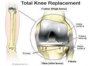
 A total knee replacement is a surgical procedure whereby the diseased knee joint is replaced with artificial material. The knee is a hinge joint which provides motion at the point where the thigh meets the lower leg. The thigh bone abuts the large bone of the lower leg at the knee joint. During a total knee replacement, the end of the femur bone is removed and replaced with a metal shell.
What is Unicondylar and Patellofemoral ?
Unicondylar and Patellofemoral is a knee replacement . unicompartmental knee arthroplasties involves placing an implant on just one side of the knee , rather than over the entire surface of the knee joint , as in a total knee replacement . refers to pain under and around the knee cap. The pain of patellofemoral may occur in one or both knees , and it tends to worsen with activity, while descending stairs and after long periods of inactivity. Patellofemoral pain syndrome is often mistaken for chondromalacia, a condition which describes damage (typically softening) of the articular cartilage on the underside of the kneecap (patella).
There are different treatments depending on condition which Dr.Kevin yip will advise during the consultation, if there is a need for a treatment or surgery. For an appointment with Dr. Kevin yip please call +65 6471 2691(24hours).
Total Knee Replacement Surgery
The patient is first taken into the operating room and given anesthesia. After the anesthesia has taken effect, the skin around the knee is thoroughly scrubbed with an antiseptic liquid. The knee is flexed about 90 degrees and the lower portion of the leg, including the foot, is placed in a special device to securely hold it in place during the surgery. Usually a tourniquet is then applied to the upper portion of the leg to help slow the flow of blood during the surgery. An incision of appropriate size is then made.
Removing the Damaged Bone Surfaces
The damaged bone surfaces and cartilage are then removed by the surgeon. Precision instruments and guides are used to help make sure the cuts are made at the correct angles so the bones will align properly after the new surfaces (implants) are attached.
Small amounts of the bone surface are removed from the front, end, and back of the femur. This shapes the bone so the implants will fit properly.The amount of bone that is removed depends on the amount of bone that has been damaged by the arthritis.
A small portion of the top surface of the tibia is also removed, making the end of the bone flat. The back surface of the patella (kneecap) is also removed.
Attaching the Implants
An implant is attached to each of the three bones. These implants are designed so that the knee joint will move in a way that is very similar to the way the joint moved when it was healthy. The implants are attached using a special kind of cement for bones.
The implant that fits over the end of the femur is made of metal. Its surface is rounded and very smooth, covering the front and back of the bone as well as the end.
The implant that fits over the top of the tibia usually consists of two parts. A metal baseplate fits over the part of the bone that was cut flat. A durable plastic articular surface is then attached to the baseplate to serve as a spacer between the baseplate and the metal implant that covers the end of the femur.
The implant that covers the back of the patella is also made of a durable plastic.
Artificial knee implants come in many designs. Some designs may have pegs, requiring small holes to be drilled into the bone after the damaged surfaces have been removed. Others may have central stems. In addition, some designs may allow screws to be used on the lower implant to provide added attachment security. The surgeon will choose the implant design that best meets the patient’s needs.
Closing the Wound
If necessary, the surgeon may adjust the ligaments that surround the knee to achieve the best possible knee function.
When all of the implants are in place and the ligaments are properly adjusted, the surgeon sews the layers of tissue back into their proper position. A plastic tube may be inserted into the wound to allow liquids to drain from the site during the first few hours after surgery. The edges of the skin are then sewn together, and the knee is wrapped in a sterile bandage. The patient is then taken to the recovery room.
